Dietary treatments for major poultry diseases
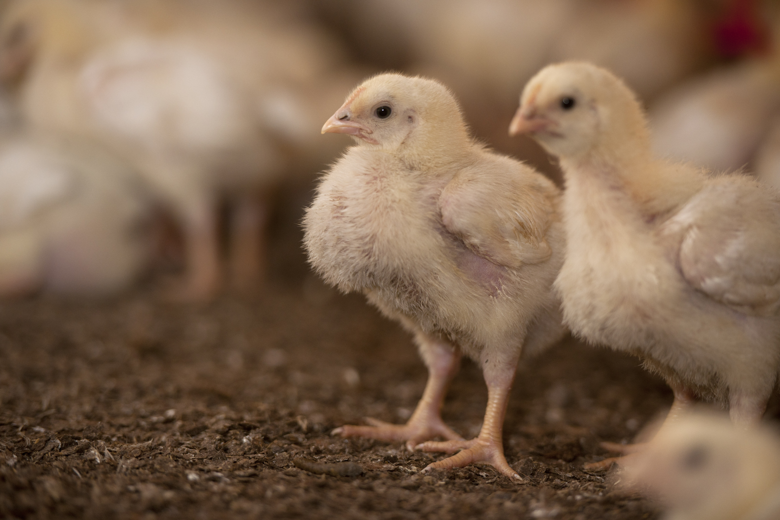
Necrotic enteritis and coccidiosis are important diseases that affect the poultry industry worldwide. Butyrate has a multifactorial role in intestinal health and can thus be a solution to manage these diseases. Here we describe some of the latest insights and studies performed on this topic.
In the poultry industry, the difference between making a profit or loss is often determined by the incidence of infectious diseases. Coccidiosis and necrotic enteritis are important concerns globally due to production losses, increased mortality, increased veterinarian and medication costs, the reduced welfare of birds and an increased chance of contamination in products for human consumption. Although both diseases have a different pathology, they act synergistically because the development of necrotic enteritis is highly dependent on the intestinal damage caused by coccidiosis. Anti-coccidial drugs and conventional antimicrobial agents can be used to control the pathogenic pressure in a flock. But the emergence of drug-resistant strains, especially after a prolonged use of drugs, is a real problem. The poultry industry therefore needs to search for alternative management and dietary strategies to control coccidiosis and necrotic enteritis. One of the dietary treatments could be the addition of butyrate, an energy source for epithelial cells that reinforces the intestinal barrier function.
Avian coccidiosis
Coccidiosis in poultry is caused by a parasitic protozoan Eimeria. The main concern from coccidiosis is that disease is not related to a single Eimeria spp. Infections result from a mixture of Eimeria species that invade different parts of the intestine, as shown in Figure 1. The intracellular parasite invades and destroys the epithelial cells of the host, causing severe damage to the intestinal wall. Nine different Eimeria species are known in poultry, only five to seven of which are associated with diseases in commercial flocks. Whereas an E. praecox infection is generally considered to produce little pathology, an infection with E. acervulina and E. mitis can result in mild enteritis followed by fluid loss and the malabsorption of nutrients. In more severe cases, inflammation of the intestinal wall with local bleeding (haemorrhages) and sloughing of epithelia (E. brunetti, E. maxima) or complete villar destruction resulting in extensive haemorrhage and death (E. necatrix, E. tenella) is seen in infected chickens1,2. Most of the highly pathogenic species invade the lower parts of the digestive tract. The life cycle of Eimeria is relatively short, from four to six days, and consists of two developmental stages; exogenous (in faecal matter) and endogenous (in host digestive tract). The exogenous stage starts after the release of unsporulated (non-infective) oocysts in the faeces. Sporulation of the oocyst occurs in the faeces and is encouraged by the right temperature, humidity and aeration (access to oxygen). In the sporulated oocyst, so-called sporozoites are formed and, from that point on, the oocysts are considered infectious. Sporulated oocysts are remarkably hard and protect the parasite from desiccation and chemical disinfectants, ensuring long-term survival in a poultry house environment1,2,3. The endogenous stage starts after ingestion of the infective oocysts by the chicken. In the micro-environment of the gizzard, sporozoites are released from the oocyst. Further down the digestive tract, the sporozoites invade and destroy epithelial cells and start the highly efficient reproduction cycle. This involves several rounds of asexual reproduction, followed by sexual differentiation, fertilisation and the shedding of unsporulated oocysts. The highly efficient copious reproduction of Eimeria species in the digestive tract and perfect survival strategy (sporulation) in faecal matter increase the chances of infection in avian flocks2.
Vaccinations and dietary treatments
Good husbandry helps to reduce the risk of transmitting the coccidiosis-causing parasites. Additional treatments are
essential, especially during the more delicate endogenous stage in the host digestive tract. Two categories of anticoccidial drugs are applied, i.e. ionophorous compounds (ionophores) and synthetic drugs (chemicals). Generally, ionophores cause death (coccidiocidal) to the parasite by interfering with the passage of ions across the cell membrane, whereas chemicals prevent the replication and growth (coccidiostatic) by inhibiting different biochemical pathways of the parasite1. To reduce the emergence of drug-resistant strains, shuttle and rotation programmes are widely used2. Still, the demand for alternative methods, such as vaccinations and dietary treatments, is increasing due to pressure from governmental agencies and consumers to ban the use of drugs in animals intended for human consumption. Moreover, coccidiosis resistance to anticoccidials leads to economic losses for the industry.
Use of vaccines
Besides the number of ingested sporulated oocysts, the severity of coccidiosis highly depends on there being immunological memory for the pathogen. As early as 1923, Johnson published the first articles showing that resistance to a dose of oocysts was not age-dependent, but relied on earlier exposure to the parasite2. Today, we still make use of this knowledge through the application of live (attenuated) vaccines. From a dietary point of view, different strategies can be used to fight coccidiosis. Some products act anti-microbially against Eimeria-specific species, e.g. essential oils and herbal extracts. Other products beneficially modulate the immune status of the chicken, whereas prebiotics and probiotics improve microflora to reduce the chance of secondary infections (e.g. C. perfringens). Damaged intestinal tissue benefits from additional antioxidants to reduce the vicious cycle of oxidative stress caused by damaged cells1. Other products improve intestinal protection and mucosal healing, e.g. betaine, butyrate or threonine4,5,6.
Necrotic enteritis lesions
Coccidiosis in poultry often pre-exists or occurs concurrently with field outbreaks of necrotic enteritis7. The causative agent of necrotic enteritis is Clostridium perfringens, a gram-positive spore forming anaerobic bacteria commonly found in soil, dust, faeces, feed, poultry litter and intestinal contents8. Necrotic enteritis has long been controlled by using antibiotic growth promoters (AGPs) in feed. Nonetheless, in January 2006, the ban on dietary AGPs took effect in Europe and the use of AGPs is under discussion on other continents. Necrotic enteritis has emerged as a common broiler disease worldwide9,10. The conventional therapeutic antimicrobial agents and anti-coccidial drugs, which not only exert an effect against Eimeria spp but also against C. perfringens, are currently used to control necrotic enteritis8. But this approach conflicts with the objective of reducing the use of antibiotics in animal production. Understanding the pathology and predisposing factors of necrotic enteritis is helpful in searching for preventive alternatives. Necrotic enteritis usually occurs three to four weeks after hatching11. The necrotic lesions are mainly restricted to the small intestine and the infection can result in an acute clinical disease or is present in a subclinical condition. In clinical cases, there is increased flock mortality during the last weeks of rearing, often without premonitory signs. The disease is acute, with death occurring within one to two hours, and mortality rates can rise up to 50%. Broiler flocks with subclinical necrotic enteritis do not display clear clinical signs and usually there is no peak mortality. Chronical damage to the intestinal mucosa leads to poor digestion and absorption of nutrients, resulting in reduced weight gain and increased feed conversion9,10,12. In subclinical conditions, feed intake can be reduced by 35% during the infectious period13. In certain cases, intestinal damage can allow C. perfringens to reach the bile duct and portal blood stream. Colonisation of C. perfringens in the liver results in cholangiohepatitis, lesions to the liver, which are finally found at the slaughter line12,14. Although clinical outbreaks of necrotic enteritis may cause high levels of mortality, the subclinical form is economically more important, as it often stays undetected in the broiler flock. Hampered growth and an increased number of condemnations at the slaughter line causes significant economic losses to the poultry farmer. The true economic impact of necrotic enteritis is not from birds that die from infection, but rather those that suffer from disease and survive the subclinical form (12).
Available nutrients allow proliferation
Clostridium perfringens is commonly found in the normal healthy avian gut microbiota. The virulence of a strain depends on its nature. Strains of C. perfringens are classified into five different types (A to E) on the basis of the production of four major toxins. Clostridium type A is associated with necrotic enteritis in broilers, although this type is also found in the digestive tract of healthy poultry9. As necrotic enteritis is a multifactorial disease, more predisposing factors are needed for its development (Figure 2)10. High levels of (animal) protein or poorly digestible protein sources are associated with a higher risk of necrotic enteritis, as undigested protein is a growth substrate for pathogenic bacteria such as C. perfringens. Wheat, rye, oats and barley are more often associated with necrotic enteritis, due to higher levels of indigestible, water soluble, non-starch polysaccharides, whereas maize is not. Besides the choice of raw materials, particle size also seems to influence gut health. Feed containing many small and some large-sized particles is more predisposed to necrotic enteritis than feed containing uniform particles7,10,15. Changes in the feeding regime (moving from starter to grower diets), other diseases and increased stocking density increase stress in the flock and suppress the immunological status of the chickens, making them more sensitive to necrotic enteritis infection12. Coccidiosis infection is an important predisposing factor, as the mucosal damage caused by Eimeria provides a favourable environment for C. perfringens to proliferate. Synergism between coccidiosis-causing protozoan Eimeria and necrotic enteritis-inducing C. perfringens is shown in Figure 3. It is interesting to note that C. perfringens needs high-quality protein, as it requires 13 essential amino acids12. By killing epithelial cells, Eimeria induces leakage of plasma proteins (A) and the coccidial infection enhances mucus production (B). Both effects provide an increased nutrient availability for C. perfringens to grow12. The virulence of the C. perfringens strain depends mainly on four major factors: its ability to produce bacteriocins, bacterial collagenolytic enzymes, toxins and its ability to adhere to the gut wall (Figure 2). In the initial stage of necrotic enteritis, collagenolytic enzymes affect the extracellular matrix and cellular junctions. Both host collagenases (probably stimulated by coccidial infection) and bacterial collagenolytic enzymes may play a role in the disorganisation or even complete destruction of the cellular matrix and the tight junctions between epithelial cells (C). Production of bacteriocins by a virulent strain inhibits the growth of other C. perfringens strains in the gut. This has the maximum benefit of increased nutrient availability released by the coccidiosis infection (D)12.
NetB: A novel toxin
For a long time, α-toxin was proposed as the main virulent factor for C. perfringens-inducing necrotic enteritis. Recently, researchers described a novel toxin called NetB. This toxin seems to be involved in the induction of necrotic lesions by forming pores in epithelial cells, leading to cell death (E)9. Virulent strains of C. perfringens are capable of binding to extracellular matrix molecules (F), a survival strategy used by numerous bacterial enteropathogens. Normal healthy intestinal epithelium doesn’t expose these extracellular matrix molecules. Because of the intestinal damage induced by Eimeria (ECMMs), NetB toxins and the colleagenolytic enzymes, C. perfingens is better able to bind to and colonise in the digestive tract, resulting in even more severe lesions12. Strategies to control necrotic enteritis without the use of AGPs and prophylactic or therapeutic treatment is challenging. To date, there is no single strategy to combat C. perfringens-associated necrotic enteritis. The combination of good hygiene management in poultry houses, the use of vaccination (against C. perfringens and coccidiosis) and dietary interventions can, to some extent, be an alternative to antibiotics to maintain production and control necrotic enteritis. Low-protein diets or the use of highly digestible protein sources in combination with enzymes to break down the undigestible structural components in the diet will reduce the opportunity for C. perfingens to develop in the gut8. Also, the use of additives to control coccidiosis, as earlier mentioned in this article, is a helpful tool to reduce the incidence of necrotic enteritis.
Multifarious effect of butyrate
The constant immunological challenge presented by Eimeria species, concurrently with C. perfringens, makes the barrier defense function of the epithelial layer extremely important. Butyrate is a short-chain fatty acid that is naturally produced in the digestive tract by the fermentation of fibres. The fatty acid is considered the most important energy source for intestinal cells and has multiple beneficial effects on vital intestinal functions. Research10 and practical field experience confirm that butyrate alone or in combination with other dietary strategies reduces the incidence of necrotic enteritis. Butyrate possesses a direct antimicrobial effect on C. perfringens, but this doesn’t seem to be the main factor involved. Butyrate concentrations in the digestive tract, coming from fermentation or even in combination with supplementary sources, will not reach the level required to inhibit the development of C. perfringens directly. The positive effect of butyrate is probably more related to the effects on the avian intestinal function10. In the small intestine, butyrate promotes villi development, gut morphology and function16. Further on in the digestive tract, butyrate represents the preferred energy source for the colonic cells5,17 and is a major precursor for lipid synthesis, used for incorporation into the cell membranes18. By supporting the cell membrane structure, butyrate contributes to the maintenance of the barrier and transporter functions in the gut18. Ma et al19observed an important role of butyrate in intestinal wound-healing owing to its positive effect on tight junctions and gut integrity. At low concentrations, butyrate reinforces the intestinal defense barriers by increasing the release of protective mucins in the mucus layer20,21 and the release of antimicrobial peptides22,23,24. These peptides, also called host defense peptides (HDPs), possess a broad spectrum of antimicrobial activity against bacteria, protozoa, enveloped viruses and fungi. HDPs bind to the microbial membrane and cause membrane disruption, which results in microbial death25. The research group of van Immerseel26 shows another antimicrobial efficacy of butyrate, whereby the fatty acid reduces the ability of pathogenic bacteria to adhere to the gut cell wall. Finally, butyrate acts as an anti-inflammatory agent27,28. Tempering the inflammatory status of the chicken is a useful tool to overcome feed intake reduction and thereby reduce the breakdown of muscle tissue during necrotic infection13. Uncoated butyrate is directly absorbed in the first part of the small intestine and does not reach the lower parts of the digestive tract29. Proper coating of dietary butyrate is essential for a targeted release of the fatty acid over the whole digestive tract30,31. By testing the micro-encapsulated butyrate product Excential Butycoat, both in vivo and in vitro, it is shown that all butyrate passes the stomach and is gradually released in the intestinal tract.
Conclusion
Since the ban on in-feed antibiotics, necrotic enteritis caused by C. perfringens has re-emerged in avian flocks with a severe economic impact. As the occurrence of drug-resistant strains is a significant problem for public health and long-term control strategies against necrotic enteritis, solutions to control necrotic enteritis must be found in alternatives to conventional medication. Nutrition strongly influences the incidence of necrotic enteritis and other pre-existing pathogenic infection coccidiosis. Proper micro-encapsulated butyrate in the diet ensures a targeted release of butyrate throughout the digestive tract, thus supporting intestinal cells and reinforcing the mucosal barrier function damaged by Eimeria species and C. perfringens.
References are available on request.
You can enjoy more articles like this, by logging on or registering here for free.


