New PCR-based test replaces microscopy screening of feed
The new 2009 real-time PCR-based method will become the US Food and Drug Administration’s “one-stop shop” for testing animal feed for prohibited materials, replacing feed microscopy as the method of choice.
By Dick Ziggers
The Office of Research (OR) of the US Food and Drug Administration’s Center for Veterinary Medicine (CVM) recently developed a new method for testing animal feed for prohibited materials of mammalian origin. The method relies on polymerase chain reaction (PCR), a molecular technique that amplifies small amounts of genetic material (DNA or RNA) to produce larger amounts for analysis. Once the new PCR-based method is routinely used, it will enhance the FDA’s ability to make sure animal feed is safe and free of prohibited materials that may spread the agent thought to cause bovine spongiform encephalopathy (BSE), or “mad cow disease”.
BSE was first detected in the United Kingdom in 1986. Studies quickly established the association between outbreaks of BSE and the use of cattle feed containing protein from cattle and other ruminants, such as sheep and goats. Then, in 1988, the UK issued the world’s first feed ban prohibiting ruminant meat-and-bone meal from being fed to cattle and other ruminants.On 5 June, 1997, the FDA issued a similar feed ban that prohibited most mammalian protein from being used to make animal feed for ruminants. In April 2008, the FDA strengthened the feed ban by prohibiting high-risk materials from being used to make all animal feed, including pet food. High-risk materials are those materials from cattle that have the highest chance of carrying the agent thought to cause BSE, such as the brains and spinal cords from cattle 30 months of age or older.
Current feed sample testing
To make sure that animal feed manufacturers comply with the feed ban, the FDA tests feed samples for prohibited materials. These samples are typically collected by field investigators in the Office of Regulatory Affairs (ORA), the FDA’s investigative arm. The samples are analysed by feed microscopy, a technique that uses a microscope to visually identify the components in the sample. Samples that test positive for a prohibited mammalian protein by feed microscopy then undergo PCR testing to confirm the result.
In 2001, the FDA validated a PCR-based method capable of detecting mammalian protein in animal feed. The OR’s feed analysts used this method for several years when they were asked by the ORA to confirm the presence of prohibited materials in animal feed, although the method was never used by field investigators in the ORA. In 2006, the OR validated an improved version of the PCR-based method from 2001. After extensive hands-on training, ORA field investigators began using the 2006 method to confirm positive feed microscopy results.
Traditional PCR-based method
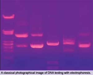
Both the 2001 and 2006 PCR-based methods rely on agarose gel electrophoresis, which is a technique that separates materials by size using an electric field applied to a gel. In agarose gel electrophoresis, the gel is made from agarose, a gelatinous substance derived from seaweed. The DNA from an animal feed sample is extracted and amplified using PCR. The DNA can be made visible by using electrophoresis, fluorescent dies, UV-light and photography, which results in the well-known DNA profile bands.
PCR-based methods that rely on UV light for visualising the bands commonlyhave problems. Most of the problems encountered during OR’s validation testing of the 2006 method had to do with the process of agarose gel electrophoresis, or interpreting the resulting photograph. For example, when a group of feed samples containing negative, slightly positive, and highly positive samples is analysed, the resulting photograph is often a hard-to-read compromise between the lowest-intensity bands, which are faint to undetectable, and the highest-intensity bands, which are so bright that they hide nearby lanes. Also, the instrument that takes the photograph of the gel is usually set to automatically focus on the brightest bands, causing bands of lesser intensity to be barely visible or not visible at all. Another problem with the 2006 method is that it takes at least 8 hours to analyse a sample.
Improvements of the 2009 method
An important criterion was the ability of the new method to detect mammalian protein when present in animal feed at a concentration of at least 0.1%. This value was chosen because:
• 0.1% was the concentration used in the validation trials for the 2001 and 2006 PCR-based methods; and
• 0.1% is the concentration that is normally detected by feed microscopy.
The new real-time PCR-based method met strict requirements for sensitivity (ability to detect true positive samples), selectivity (ability to detect true negative samples), and specificity (ability to detect only the targeted animal species). It also met strict requirements for ruggedness and real-time platform. The ruggedness test determines how well the method tolerates small changes to its set operating limits and measures the method’s reliability under normal use. The real-time platform test assesses how well the PCR portion of the method works using different laboratory instruments.A peer verification trial with two outside laboratories proved that the new real-time PCR-based method can easily and reliably be used by other laboratories.
Advantages of the 2009 method
In less than 2.5 hours, the new real-time PCR-based method can detect processed materials from cattle, sheep, and goats, as well as a select set of processed materials from chickens, turkeys, and geese. The method can also detect animal materials that have been processed in both North America and the EU. The processing conditions in the EU are very different from those in North America, resulting in meat-and-bone meals with different characteristics. Because it can detect meat-and-bone meals processed in both North America and the EU, the 2009 method is more useful and versatile.
Besides being faster and more versatile, another advantage of the new real-time PCR-based method is that all the components are available as a pair of commercial kits that are made under strict quality controls. One kit contains the reagents needed to extract the DNA from the feed sample for amplification by PCR, and the second kit contains the reagents needed to perform the PCR test. Having all the necessary reagents available for laboratories in ready-to-use kits that have already been examined for quality control further reduces the time needed to analyse a sample.
Source: AllAboutFeed vol 1 nr 1, 2010


